

Biochemie & Zellbiologie der Lipide
Prof. Dr. Christoph Thiele

LD540
- LD540: A new tool to image LDs in fixed and live cell samples
In the past, LDs have been visualized in microscopy using various lipophilic dyes. Amoung others, BODIPY and Nile Red were found to be sufficient for this specific application. However, their fluorescent spectral properties and sensitivity to photobleaching make them unsuitable in multicolor imaging, especially in combination with the commonly used GFP, and long-term live-cell imaging approaches.Therefore we developed a new lipophilic dye, called LD540 (Spand et al. 2009), which suits the demands of live cell and multicolor imaging applications by being photostable and having a fluorescent-spectrum allowing the usage of LD540 in combination with most commonly used fluorophores like GFP, DAPI and Alexa647.
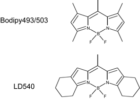
Structures of LD540 and Bodipy 493/503.
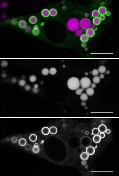
Live cell two-color imaging of 3T3-L1 cells transiently expressing AUP1-EGFP. Cells were stained with LD540 and imaged as described above. An overlay of green (AUP1-EGFP) and magenta (LDs) channels is shown together with the single unprocessed tiff files to demonstrate the complete separation of the two channels. Bar: 10 μm.
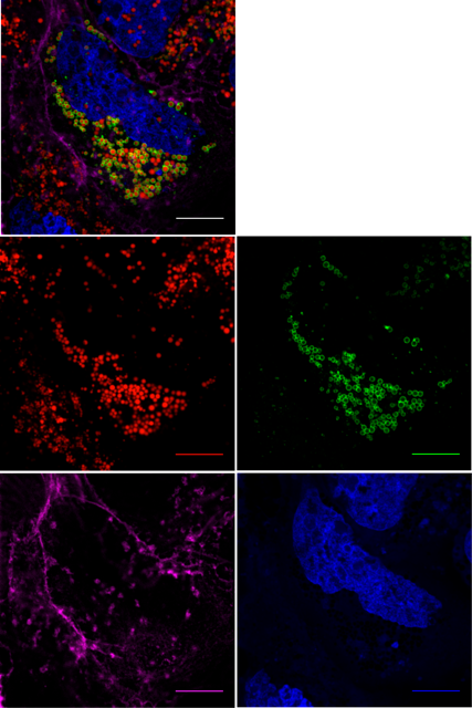
Multicolor imaging of LDs in HuH7 hepatoma cells. HuH7 cells transiently expressing Rab18-EGFP (green) were fixed, permeabilized, and stained with Alexa647-phalloidin (magenta) for actin filaments, LD540 (red) for LDs and DAPI (blue) for DNA. Bar: 10 μm.
After we originally described LD540 [II] we and others have successfully used the dye to stain LDs in various cell types [I, III].